Posts

Difinition of lungs , Anotomy,cause of lungs disorders, symptoms, functions, condition, treatment and test s
Image
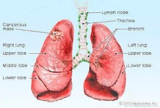
 Definition of Lung, Anatomy,Causes of lung disorders, Symptoms, Function,Conditions, Treatment and tests. The lungs are a pair of spongy, air-filled organs located on either side of the chest (thorax). The trachea (windpipe) conducts inhaled air into the lungs through its tubular branches, called bronchi. The bronchi then divide into smaller and smaller branches (bronchioles), finally becoming microscopic. The bronchioles eventually end in clusters of microscopic air sacs called alveoli. In the alveoli, oxygen from the air is absorbed into the blood. Carbon dioxide, a waste product of metabolism, travels from the blood to the alveoli, where it can be exhaled. Between the alveoli is a thin layer of cells called the interstitium, which contains blood vessels and cells that help support the alveoli. The lungs are covered by a thin tissue layer called the pleura. The same kind of thin tissue lines the inside of the chest cavity -- also called pleura. A thin layer of fluid acts as a lu...

Spleen (definition, structure, location,types, functions,disease and surgery procedures

What is Spleen? An organ that is part of the lymphatic system. The spleen makes lymphocytes, filters the blood, stores blood cells, and destroys old blood cells. It is located on the left side of the abdomen near the stomach. Structure : The spleen has a slightly oval shape. It is covered by a weak capsule that protects the organ whilst allowing it to expand in size. The outer surface of the spleen can be anatomically divided into two: 1)Diaphragmatic surface – in contact with diaphragm and ribcage. 2)Visceral surface – in contact with the other abdominal viscera. It has anterior, superior, posteromedial and inferior borders. The posteromedial and inferior borders are smooth, whilst the anterior and superior borders contain notches. In enlargement of the spleen (known as splenomegaly), the superior border moves inferomedially, and its notches can be palpated. The spleen is made up of two basic types of tissue, each with different functions: 1)White pulp 2)Red pulp 1)The white pulp is p...

Cell membrane ( कोशिका झिल्ली)

Image
 Cell membrane ( कोशिका झिल्ली) :- कोशिका के सभी  अव्यय एक झिल्ल  से गिरे रहते हैं इस झिल्ली को कोशिका झिल्ली कहते हैं कोशिका झिल्ली का निर्माण 3 परतों से मिलकर बना। इसमें बाहरी व भीतरी परतों का निर्माण प्रोटीन के द्वारा होता है मध्य  परत का  निर्माण वसा व लिपिड से होता हैं कोशिका झिल्ली कोशिका के "जीव द्रव"को घेर कर रखती है।   Protoplasm (जीव द्रव):-   कोशिका के कोशिका झिल्ली के अंदर संपूर्ण पदार्थ को जीव द्रव कहते हैं। ये दो भागों से मिलकर बना है। 1. Nucleus (केंद्रक) 2.cytoplasm(कोशिका द्रव) Nucleus(केंद्रक):- कोशिका द्रव मैं स्थित संरचना जो जीव द्रव के क्रिया को संचालित करता है या  कोशिका का नियंत्रण करता है  nucleus ( केंदक )कहलाता है। केंद्रक गोलाकार अंडाकार होता है इसका व्यास 5 माइक्रोमीटर से 20 माइक्रोमीटर होता है यह है कोशिका का सबसे बड़ा कोशिकांग होता हैं । Nucleus functions-(कार्य) 1. कोशिका के विकास व प्रजनन को नियंत्रण करता है। 2.इसलिए इसे "सेेल की लाइफ "भी कहा जाता है।   Cytoplasm(कोशिका द्रव):- कोशिका में कोशिका झिल्...

Cell (कोशिका)

Image
   Cell(कोशिका)-  सजीवो की मूलभूत संरचनात्मक और क्रियात्मक इकाई है। यह शरीर का छोटा से छोटा संगठित रूप है‌‌। जिस से सभी कि्या होती हैं। कोशिका का अध्ययन" कोशिका विज्ञान" या "CYTOLOGY" कहलाता हैं। कोशिका की खोज "रोबर्ट हुक" ने   "1665" में की जिंदा कोशिका (live cell) की खोज " antonvan Leeuwenhoek" ने "1674"    की। Diagram-

Cell(कोशिका)

 Cell( कोशिका )-सजीवो की मूलभूत सर्चनातमक और क्रियात्मक इकाई है यह शरीर का छोटा से छोटा संगठित रूप है‌‌।जिससे शरीर की सभी क्रियाएं होती हैं। कोशिका का अध्ययन "कोशिका विज्ञान" कहलाती हैं। कोशिका की खोज"रोबर्ट हुक" ने "1665 " में की। ज़िन्दा कोशिका की खोज " van Leeuwenhoek " ने"1674" में की।